| CAS Number | 569-61-9 |
|---|---|
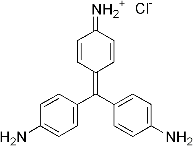
| Molecular Formula | C19H18ClN3 |
| Molecular Weight | 323.824 |
| InChI Key | JUQPZRLQQYSMEQ-UHFFFAOYSA-N |
| Synonyms |
|
Applications:
UV-Vis Spectrum of Pararosaniline
July 16, 2024
Access the UV-Vis Spectrum SIELC Library

If you are looking for optimized HPLC method to analyze Pararosaniline Hydrochloride check our HPLC Applications library
For optimal results in HPLC analysis, it is recommended to measure absorbance at a wavelength that matches the absorption maximum of the compound(s) being analyzed. The UV spectrum shown can assist in selecting an appropriate wavelength for your analysis. Please note that certain mobile phases and buffers may block wavelengths below 230 nm, rendering absorbance measurement at these wavelengths ineffective. If detection below 230 nm is required, it is recommended to use acetonitrile and water as low UV-transparent mobile phases, with phosphoric acid and its salts, sulfuric acid, and TFA as buffers.
For some compounds, the UV-Vis Spectrum is affected by the pH of the mobile phase. The spectra presented here are measured with an acidic mobile phase that has a pH of 3 or lower.

HPLC Method for Analysis of Pararosaniline, Crystal Violet, and Crystal Violet Lactone on Primesep 100 Column
December 7, 2022
High Performance Liquid Chromatography (HPLC) Method for Analysis of Crystal Violet, Crystal Violet Lactone, Pararosaniline Hydrochloride on Primesep 100 by SIELC Technologies.
Pararosaniline (Basic Red 9) is a popular basic magenta dye and part of the triarylmethane family of dyes with the chemical formula C19H17N3. It is a free base version of pararosaniline hydrochloride. Primarily, it is used to dye synthetic materials, to detect sulfur dioxide, and as an antischistosomal. You can find detailed UV spectra of Pararosaniline and information about its various lambda maxima by visiting the following link.
Crystal Violet (Methyl Violet 10B), another basic triarylmethane dye, has the C25H30ClN3. It is frequently used for histological stains and for identifying Gram-positive bacteria. It is said to have antibacterial, antifungal, and anthelmintic properties. It is a common component of navy blue and black inks in printing, inkjet printers, and ball-point pens. You can find detailed UV spectra of Crystal Violet and information about its various lambda maxima by visiting the following link.
Crystal Violet Lactone, a derivative of Crystal Violet, is a basic thermochromic dye with the chemical formula C26H29N3O2. It is widely used as a security marker for various types of fuels as well as carbonless copy papers. It is a slightly yellow crystalline powder that is soluble in nonpolar organic solvents.
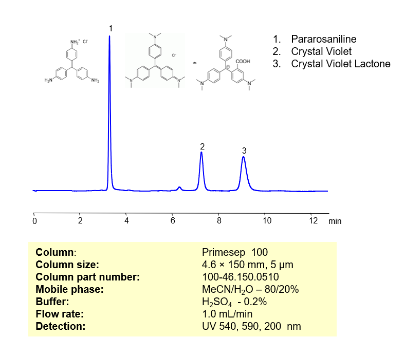
These three basic dyes can be separated, retained, and analyzed on a Primesep 100 mixed-mode stationary phase column using an isocratic analytical method with a simple mobile phase of water, Acetonitrile (MeCN), and a sulfuric acid (H2SO4) buffer. This analysis method can be detected in the UV-Vis regime at 540, 590, and 200 nm.
Condition
| Column | Primesep 100, 4.6 x 150 mm, 5 µm, 100 A, dual ended |
| Mobile Phase | MeCN/H2O – 80/20% |
| Buffer | H3PO4 – 0.2% |
| Flow Rate | 1.0 ml/min |
| Detection | UV, 540, 590, 200 nm |
| Peak Retention Time | 3.15, 7.24, 8,82 min |
Description
| Class of Compounds | Dyes |
| Analyzing Compounds | Crystal Violet, Crystal Violet Lactone, Pararosaniline Hydrochloride |
Application Column

Primesep 100
Column Diameter: 4.6 mm
Column Length: 150 mm
Particle Size: 5 µm
Pore Size: 100 A
Column options: dual ended
Crystal Violet Lactone
Pararosaniline Hydrochloride
HPLC Method for Analysis of Pararosaniline and Ethyl Red on Primesep 100 Column
December 7, 2022
High Performance Liquid Chromatography (HPLC) Method for Analysis of Pararosaniline and Ethyl Red on Primesep 100 by SIELC Technologies.
Pararosaniline (Basic Red 9) is a popular basic magenta dye and part of the triarylmethane family of dyes with the chemical formula C19H17N3. It is a free base version of pararosaniline hydrochloride. Primarily, it is used to dye synthetic materials, to detect sulfur dioxide, and as an antischistosomal. You can find detailed UV spectra of Pararosaniline and information about its various lambda maxima by visiting the following link.
Ethyl Red is a pH indicator with C₁₇H₁₉N₃O₂ as its molecular structure. When the pH transitions from acidic to neutral, Ethyl red turns from yellow to red, hence the name. Outside of experiments, it is occasionally used as a dye in textiles and foods. You can find detailed UV spectra of Pararosaniline Hydrochloride, Ethyl red and information about its various lambda maxima by visiting the following link.
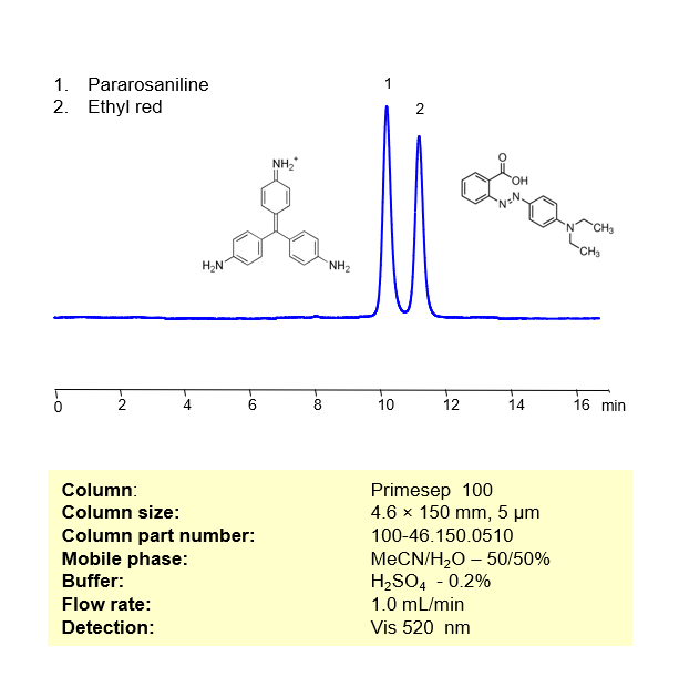
These two basic dyes can be separated, retained, and analyzed on a Primesep 100 mixed-mode stationary phase column using an isocratic analytical method with a simple mobile phase of water, Acetonitrile (MeCN), and a sulfuric acid (H2SO4) buffer. This analysis method can be detected in the visible regime at 520 nm.
Condition
| Column | Primesep 100, 4.6 x 150 mm, 5 µm, 100 A, dual ended |
| Mobile Phase | MeCN/H2O – 50/50% |
| Buffer | H3PO4 – 0.2% |
| Flow Rate | 1.0 ml/min |
| Detection | UV, 520 nm |
| Peak Retention Time | 10.82 min |
Description
| Class of Compounds | Dyes |
| Analyzing Compounds | Pararosaniline Hydrochloride, Ethyl red |
Application Column
Primesep 100
Column Diameter: 4.6 mm
Column Length: 150 mm
Particle Size: 5 µm
Pore Size: 100 A
Column options: dual ended
Pararosaniline Hydrochloride
HPLC Method for Analysis of Dyes
March 1, 2018
HPLC Method for Methyl Red, Alizarin, Eosin Y, Fluorescein, Nile Blue A, Patent Blue Violet (Patent Blue), Resazurin, Carmine, Pararosaniline Hydrochloride, 3-Aminoacridine, Fluorescein Disodium Salt on Newcrom R1 by SIELC Technologies
 High Performance Liquid Chromatography (HPLC) Method for Analysis of Methyl Red, Alizarin, Eosin Y, Fluorescein, Nile Blue A, Patent Blue Violet (Patent Blue), Resazurin, Carmine, Pararosaniline Hydrochloride, 3-Aminoacridine, Fluorescein Disodium Salt
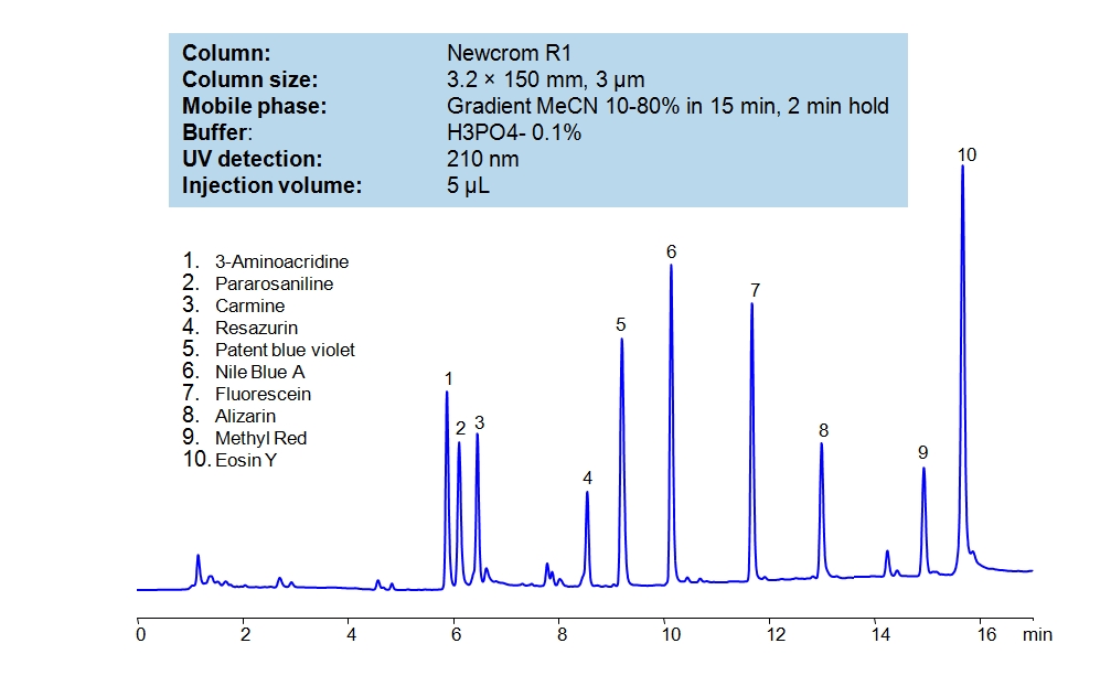
Alizarin a red pigment that comes as a red powder with a slight orange tint. When dissolved it appears bright red with a slight pink undertone. Alizarin is also known as Mordant Red 11 or Turkey Red. Eosin Y is a pigment that comes as an orange powder that when dissolved in water turns bright orange. Eosin is a fluorescent staining agent. It is used to stain red blood cells, proteins on cytoplasm, collage, and muscle fibers. It is also fluorescence. Fluorescein is a dye that comes in a dark orange powder and is used as a tracer and an indicator. As it says in the name, it is also Fluorescent, obtaining a green-yellow color when under black light. Methyl red an indicator that turns red in acidic solutions (below 4.4 pH), is a shade of orange between 4.5 and 6.1 pH, and turns bright yellow in basic solutions (above pH 6.2). Nile blue, also known as Nile blue A, is used as a stain used in biology and histology as well as a pH indicator. In pH below 7 it becomes lighter shades of glue, and at pH 0 becomes almost completely translucent. At pH of seven it has a dark blue shade, while at pH higher than that it starts becoming more red. Pararosaniline Hydrochloride, also known as Basic Red 9, or C.I. 42500. It is a a magenta solid that is used as a dye. Patent Blue Violet, also known as Patent Blue, is a deep blue pigment that is often used to dye clothes. Resazurin is a blue dye as well as a pH indicator. At pH 3.8 or lower, it is bright orange, but the higher the pH the more blue it becomes, the color stops changing at the pH of 6.5 where it becomes indigo. Newcrom R1, a column that takes advantage of the newest technologies, does not contain embedded acidic nor basic ionizable groups and can retain dyes. The method is UV compatible and can be used as a general approach for analyzing similar compounds.
| Column | Newcrom R1, 3.2 x 150 mm, 3 µm, 100 A, dual ended |
| Mobile Phase | Gradient MeCN – 10-80% |
| Buffer | Phosphoric Acid |
| Flow Rate | 0.5 ml/min |
| Detection | UV 210 nm |
| Class of Compounds | Pigments |
| Analyzing Compounds | Methyl Red, Alizarin, Eosin Y, Fluorescein, Nile Blue A, Patent Blue Violet (Patent Blue), Resazurin, Carmine, Pararosaniline Hydrochloride, 3-Aminoacridine, Fluorescein Disodium Salt |
Application Column
Newcrom R1
Column Diameter: 3.2 mm
Column Length: 150 mm
Particle Size: 3 µm
Pore Size: 100 A
Column options: dual ended
Alizarin
Carmine
Eosin Y
Fluorescein
Fluorescein Disodium Salt
Methyl Red
Nile Blue A
Pararosaniline Hydrochloride
Patent Blue Violet (Patent Blue)
Resazurin