| CAS Number | 548-62-9 |
|---|---|
| Molecular Formula | C25H30ClN3 |
| Molecular Weight | 407.990 |
| InChI Key | ZXJXZNDDNMQXFV-UHFFFAOYSA-M |
| LogP | 0.96 |
| Synonyms |
|
Applications:
UV-Vis Spectrum of Crystal Violet
July 16, 2024
Access the UV-Vis Spectrum SIELC Library
If you are looking for optimized HPLC method to analyze Crystal Violet check our HPLC Applications library
For optimal results in HPLC analysis, it is recommended to measure absorbance at a wavelength that matches the absorption maximum of the compound(s) being analyzed. The UV spectrum shown can assist in selecting an appropriate wavelength for your analysis. Please note that certain mobile phases and buffers may block wavelengths below 230 nm, rendering absorbance measurement at these wavelengths ineffective. If detection below 230 nm is required, it is recommended to use acetonitrile and water as low UV-transparent mobile phases, with phosphoric acid and its salts, sulfuric acid, and TFA as buffers.
For some compounds, the UV-Vis Spectrum is affected by the pH of the mobile phase. The spectra presented here are measured with an acidic mobile phase that has a pH of 3 or lower.

HPLC Method for Separation of Michler’s ketone and Crystal Violet on Primesep 100 Column
August 18, 2023
High Performance Liquid Chromatography (HPLC) Method for Analysis of Michler’s ketone, Crystal Violet on Primesep 100 by SIELC Technologies.
Separation type: Liquid Chromatography Mixed-mode
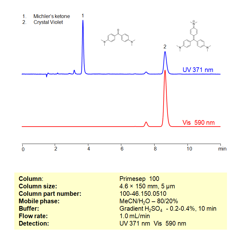
Michler’s ketone (4,4′-bis(dimethylamino)benzophenone) is a chemical compound used primarily in the synthesis of dyes and pigments. It has the chemical formula C17H17N3O2. The compound is named after the German chemist Viktor Michler, who first prepared it.
Michler’s ketone is synthesized by condensing two equivalents of dimethylaniline with one equivalent of phosgene. The reaction involves the formation of a carbonyl group (C=O) that links two dimethylaniline moieties. The resulting structure features two aromatic rings (benzene rings) connected by a carbonyl group, with dimethylamino groups attached to each aromatic ring.
Michler’s ketone is a valuable intermediate in the dye industry, where it is used to prepare various triarylmethane dyes, including Malachite Green, Methyl Violet, and Crystal Violet. The strong electron-donating properties of the dimethylamino groups make it a useful compound for synthesizing dyes with vibrant and stable colors.
Michler’s ketone and Crystal Violet can be separated, retained, and analyzed on a Primesep 100 mixed-mode stationary phase column using an isocratic analytical method with a simple mobile phase of water, Acetonitrile (MeCN), and a sulfuric acid (H2SO4) buffer. This analysis method can be detected in the UV-Vis regime at 540, 590, and 200 nm.
High Performance Liquid Chromatography (HPLC) Method for Analyses of Michler’s ketone, Crystal Violet
Condition
| Column | Primesep 100, 4.6 x 150 mm, 5 µm, 100 A, dual ended |
| Mobile Phase | MeCN/H2O – 80/20% |
| Buffer | Gr H3PO4 – 0.2-0.4%, 10 min |
| Flow Rate | 1.0 ml/min |
| Detection | UV 371, Vis 590 nm |
| Peak Retention Time | 3.71, 8.57 min |
Description
| Class of Compounds | Dyes |
| Analyzing Compounds | Michler’s ketone, Crystal Violet |
Application Column
Primesep 100
Column Diameter: 4.6 mm
Column Length: 150 mm
Particle Size: 5 µm
Pore Size: 100 A
Column options: dual ended
Michler’s ketone

HPLC Method for Analysis of Crystal Violet on Primesep 100 Column
August 17, 2023
High Performance Liquid Chromatography (HPLC) Method for Analysis of Crystal Violet on Primesep 100 by SIELC Technologies.
Separation type: Liquid Chromatography Mixed-mode
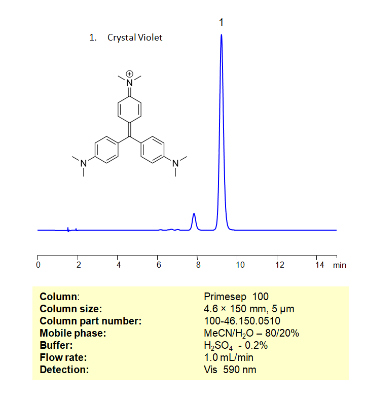
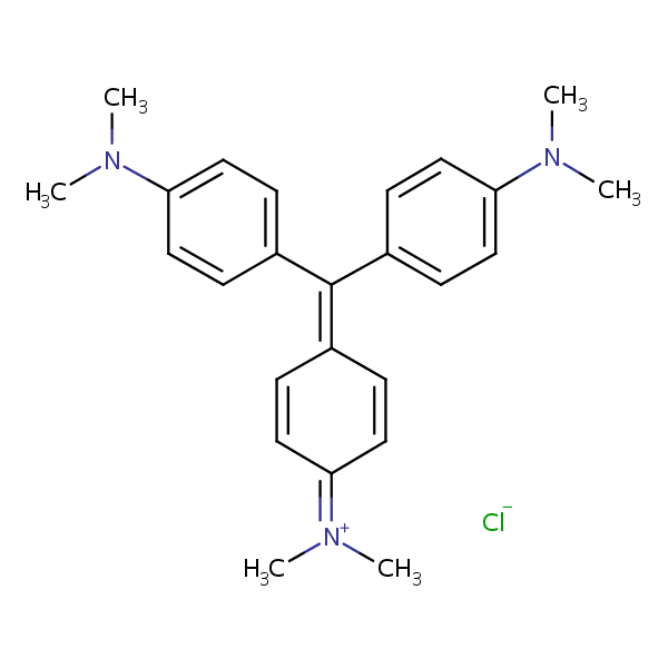
Crystal Violet, also known as Methyl Violet 10B, is a synthetic dye belonging to the triarylmethane dye family. It has the molecular formula C25H30ClN3. This dye is primarily known for its use in microbiology as a stain, but it has other applications as well.
- Gram Staining: One of the most well-known uses of Crystal Violet is in Gram staining, a method used to differentiate bacterial species into two large groups (Gram-positive and Gram-negative) based on the chemical and physical properties of their cell walls. In this process, a crystal violet solution is applied to a smear of bacterial cells, which are then treated with iodine, followed by an alcohol wash and counterstain with safranin. Gram-positive bacteria retain the crystal violet stain, while Gram-negative bacteria do not.
- Antiseptic: Crystal Violet has antiseptic properties and has been used as a topical antiseptic to treat minor burns, infections, and fungal conditions. However, due to concerns over toxicity, its use in this application has declined.
- Cell Biology and Histology: In cell biology and histology, Crystal Violet is used to stain cell nuclei, allowing for visualization under a microscope. It is also used in the assessment of cell viability and proliferation in various assays, such as the crystal violet cell cytotoxicity assay.
- Indicator in Chemistry: In analytical chemistry, Crystal Violet is used as an indicator in colorimetric titrations due to its ability to change color at different pH levels.
- Textile and Paper Industry: Crystal Violet is also used as a dye in the textile and paper industry.
Crystal Violet can be retained, and analyzed on a Primesep 100 mixed-mode stationary phase column using an isocratic analytical method with a simple mobile phase of water, Acetonitrile (MeCN), and a sulfuric acid (H2SO4) buffer. This analysis method can be detected in the UV-Vis regime at 540, 590, and 200 nm.
High Performance Liquid Chromatography (HPLC) Method for Analyses of Crystal Violet
Condition
| Column | Primesep 100, 4.6 x 150 mm, 5 µm, 100 A, dual ended |
| Mobile Phase | MeCN/H2O – 80/20% |
| Buffer | H3PO4 – 0.2% |
| Flow Rate | 1.0 ml/min |
| Detection | Vis, 590 nm |
| Peak Retention Time | 9.37 min |
| Sample concentration | 0.0006 mg/ml |
| Injection volume | 5 µl |
| LOD | 10 ppb |
Description
| Class of Compounds | Dyes |
| Analyzing Compounds | Crystal Violet |
Application Column
Primesep 100
Column Diameter: 4.6 mm
Column Length: 150 mm
Particle Size: 5 µm
Pore Size: 100 A
Column options: dual ended

HPLC Method for Analysis of Pararosaniline, Crystal Violet, and Crystal Violet Lactone on Primesep 100 Column
December 7, 2022
High Performance Liquid Chromatography (HPLC) Method for Analysis of Crystal Violet, Crystal Violet Lactone, Pararosaniline Hydrochloride on Primesep 100 by SIELC Technologies.
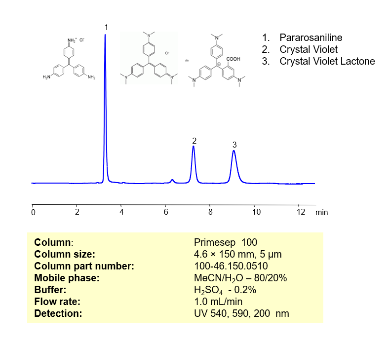
Pararosaniline (Basic Red 9) is a popular basic magenta dye and part of the triarylmethane family of dyes with the chemical formula C19H17N3. It is a free base version of pararosaniline hydrochloride. Primarily, it is used to dye synthetic materials, to detect sulfur dioxide, and as an antischistosomal. You can find detailed UV spectra of Pararosaniline and information about its various lambda maxima by visiting the following link.
Crystal Violet (Methyl Violet 10B), another basic triarylmethane dye, has the C25H30ClN3. It is frequently used for histological stains and for identifying Gram-positive bacteria. It is said to have antibacterial, antifungal, and anthelmintic properties. It is a common component of navy blue and black inks in printing, inkjet printers, and ball-point pens. You can find detailed UV spectra of Crystal Violet and information about its various lambda maxima by visiting the following link.
Crystal Violet Lactone, a derivative of Crystal Violet, is a basic thermochromic dye with the chemical formula C26H29N3O2. It is widely used as a security marker for various types of fuels as well as carbonless copy papers. It is a slightly yellow crystalline powder that is soluble in nonpolar organic solvents.
These three basic dyes can be separated, retained, and analyzed on a Primesep 100 mixed-mode stationary phase column using an isocratic analytical method with a simple mobile phase of water, Acetonitrile (MeCN), and a sulfuric acid (H2SO4) buffer. This analysis method can be detected in the UV-Vis regime at 540, 590, and 200 nm.
Condition
| Column | Primesep 100, 4.6 x 150 mm, 5 µm, 100 A, dual ended |
| Mobile Phase | MeCN/H2O – 80/20% |
| Buffer | H3PO4 – 0.2% |
| Flow Rate | 1.0 ml/min |
| Detection | UV, 540, 590, 200 nm |
| Peak Retention Time | 3.15, 7.24, 8,82 min |
Description
| Class of Compounds | Dyes |
| Analyzing Compounds | Crystal Violet, Crystal Violet Lactone, Pararosaniline Hydrochloride |
Application Column
Primesep 100
Column Diameter: 4.6 mm
Column Length: 150 mm
Particle Size: 5 µm
Pore Size: 100 A
Column options: dual ended
Crystal Violet Lactone
Pararosaniline Hydrochloride

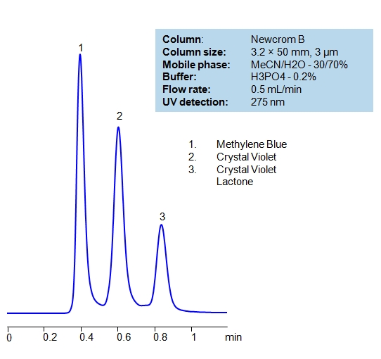
HPLC Separation of Dyes
November 21, 2010
| Column | Newcrom B, 3.2×50 mm, 3 µm, 100A |
| Mobile Phase | MeCN/H2O – 30/70% |
| Buffer | H3PO4 – 0.2% |
| Flow Rate | 0.5 ml/min |
| Detection | UV, 275 nm |
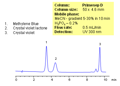
Various dyes are widely used in food, textile, and pharmaceutical industries. Dies are aromatic compounds containing ionizable groups. Methylene blue, crystal violet lactone, and crystal violet were separated on a Primesep D HPLC column. Method can be used for separation of other dyes and pigments that are basic and hydrophobic in nature.
| Column | Primesep D, 4.6×50 mm, 3 µm, 100A |
| Mobile Phase | MeCN |
| Buffer | H3PO4 – 0.2% |
| Flow Rate | 0.5 ml/min |
| Detection | UV, 300 nm |
| Class of Compounds |
Dye, Hydrophilic, Ionizable |
| Analyzing Compounds | Methylene Blue, Crystal violet lactone, Crystal violet |
Application Column
Newcrom B
The Newcrom columns are a family of reverse-phase-based columns. Newcrom A, AH, B, and BH are all mixed-mode columns with either positive or negative ion-pairing groups attached to either short (25 Å) or long (100 Å) ligand chains. Newcrom R1 is a special reverse-phase column with low silanol activity.
Select optionsPrimesep D
The Primesep family of mixed-mode columns offers a wide variety of stationary phases, boasting unprecedented selectivity in the separation of a broad array of chemical compounds across multiple applications. Corresponding Primesep guard columns, available with all stationary phases, do not require holders. SIELC provides a method development service available to all customers. Inquire about our specially-tailored custom LC-phases for specific separations.
Select optionsCrystal Violet Lactone
Methylene Blue

