
| CAS Number | 65-46-3 |
|---|---|
| Molecular Formula | C9H13N3O5 |
| Molecular Weight | 243.219 |
| InChI Key | UHDGCWIWMRVCDJ-XVFCMESISA-N |
| LogP | -2.33 |
| Synonyms |
|
Applications:
HPLC Method for Analysis of Five Nucleosides on Chromni Column
August 17, 2026
HPLC Method for Analysis of Guanosine, Cytidine, Adenosine, Uridine, Thymidine on Chromni Column by SIELC Technologies
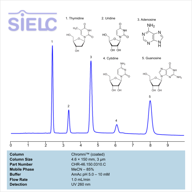
High Performance Liquid Chromatography (HPLC) Method for Analysis of Guanosine, Cytidine, Adenosine, Uridine, Thymidine.
Thymidine, also noted as dT and dThd and known as deoxythymidine, has the chemical formula C10H14N2O5. It is a a pyrimidine deoxynucleoside found as one of the four nucleosides in DNA. It pairs with deoxyadenosine in double sided DNA and is used to synchronize the cells in G1/early S phase. Thymidine’s primary use is in the production of the antiretroviral drug known as azidothymidine. You can find detailed UV spectra of Thymine and information about its various lambda maxima by visiting the following link.
Uridine, also noted as U and Urd, has C9H12O6 chemical formula. It is a pyrimidine analog containing uracil. It is one of the five standard nucleosides. It is a non-essential nutrient, since the body produces it as necessary. While uridine can be found in certain foods in the form of RNA, it is not bioavailable, as it is destroyed in the liver. You can find detailed UV spectra of Uracil and information about its various lambda maxima by visiting the following link.
Adenosine is a key building block of energy-carrying molecules with the chemical formula C10H13N5O4. It has a variety of other uses, including being a inhibitory neurotransmitter which helps with sleep and acting as a blood flow regulator. Medicinally, it is used as treatment for supraventricular tachycardia (SVT). You can find detailed UV spectra of Adenosine and information about its various lambda maxima by visiting the following link.
Cytidine, also noted as C or Cyd, is a nucleoside molecule with the chemical formula C9H13N3O5. It is primarily found in foods with high RNA contents, such as organ meets, brewer’s yeast, and beer. During digestion, Cyd is broken down into ribosyl pyrimidines. When consumed by humans, it is converted into uridine, which is suspected to be the reason behind cytidine’s metabolic effects. You can find detailed UV spectra of Cytidine and information about its various lambda maxima by visiting the following link.
Guanosine, often denoted as G or Guo, is a purine nucleoside with the chemical formula C10H13N5O5. It can be phosphorylated into many other forms, which play vital roles in biochemical possesses like synthesis of nucleic acids, proteins, photosynthesis, and more. It is also required for RNA splicing. It is used in acyclovir, an antiviral drug, to treat herpes. It can also be found in abacavir, an anti-HIV drug, and regadenoson, an A2A adenosine receptor agonist. You can find detailed UV spectra of Guanosine and information about its various lambda maxima by visiting the following link.
Guanosine, Cytidine, Adenosine, Uridine, Thymidine can be retained and analyzed using the Chromni stationary phase column. The analysis utilizes a gradient method with a simple mobile phase consisting of water and acetonitrile (MeCN) and ethanol. Detection is performed using UV.
Condition
| Column | Chromni, 4.6 x 150 mm, 3 µm, 100 A, surface coated |
| Mobile Phase | MeCN/H2O -85% |
| Buffer | AmAc pH 5.0 – 10 mM |
| Flow Rate | 1.0 ml/min |
| Detection | UV 260 nm |
Description
Application Column
Chromni
Column Diameter: 4.6 mm
Column Length: 150 mm
Particle Size: 3 µm
Pore Size: 100 A
Column options: surface coated
Cytidine
Guanosine
Thymidine
Uridine

Uv-Vis Spectrum of Cytidine
February 2, 2026
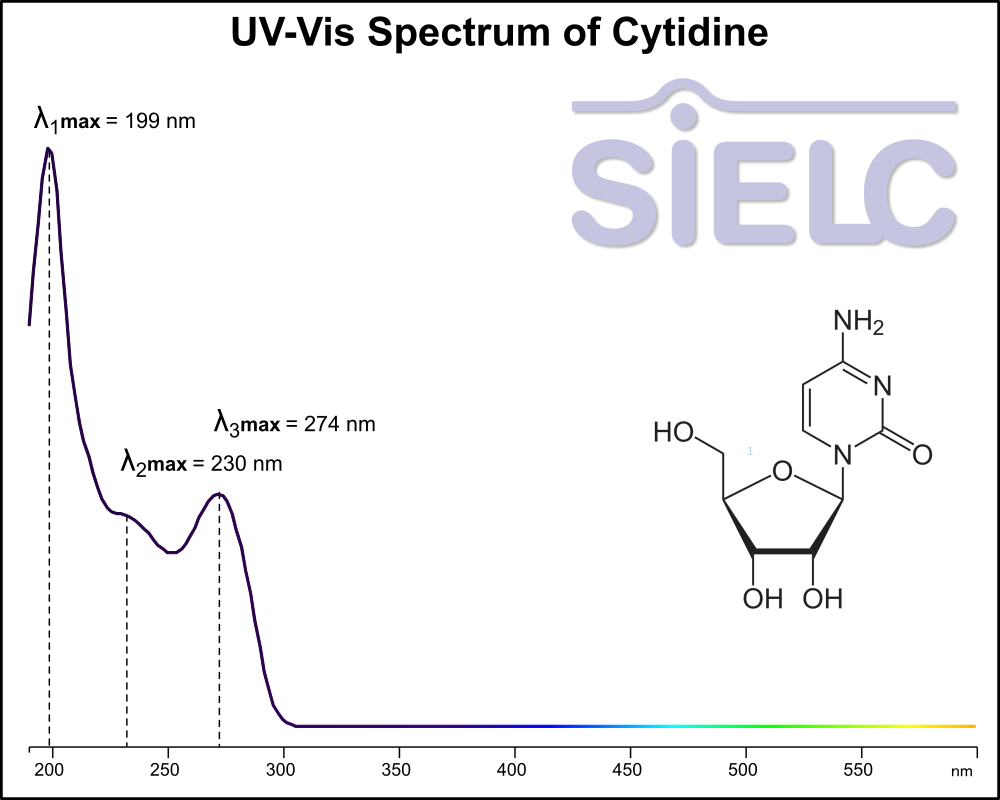
If you are looking for optimized HPLC method to analyze Cytidine check our HPLC Applications library
For optimal results in HPLC analysis, it is recommended to measure absorbance at a wavelength that matches the absorption maximum of the compound(s) being analyzed. The UV spectrum shown can assist in selecting an appropriate wavelength for your analysis. Please note that certain mobile phases and buffers may block wavelengths below 230 nm, rendering absorbance measurement at these wavelengths ineffective. If detection below 230 nm is required, it is recommended to use acetonitrile and water as low UV-transparent mobile phases, with phosphoric acid and its salts, sulfuric acid, and TFA as buffers.
For some compounds, the UV-Vis Spectrum is affected by the pH of the mobile phase. The spectra presented here are measured with an acidic mobile phase that has a pH of 3 or lower.
HPLC Method for Separation of Cytosine, Deoxycytidine and Cytidine on BIST B+ Column
November 28, 2022
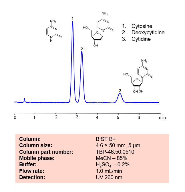
HPLC Method for Separation of Cytosine, Deoxycytidine, Cytidine on BIST B+ by SIELC Technologies.
Cytosine, also noted as C and Cyt, has the chemical formula C4H5N3O. In DNA, it pairs with Guanine to create a base pair. In RNA, it is synonymous with Uracil, being an interchangeable third base. Not only that, due to it’s instability, it can change into Uracil through spontaneous deamination.
Deoxycytidine is a deoxyribonucleoside with the chemical formula C9H13N3O4. It is a precursor for 5-aza-2′-cytidine, which is a treatment for myelodysplastic syndrome. It works through interfering with the methylation of the P15/INK4B gene. It can also be used as a biomarker for tumor diagnosis.
Cytidine, also noted as C or Cyd, is a nucleoside molecule with the chemical formula C9H13N3O5. It is primarily found in foods with high RNA contents, such as organ meets, brewer’s yeast, and beer. During digestion, Cyd is broken down into ribosyl pyrimidines.
Cytosine, Deoxycytidine, Cytidine can be retained and analyzed using the BIST B+ stationary phase column. The analysis utilizes an isocratic method with a simple mobile phase consisting of water and acetonitrile (MeCN) with a sulfuric acid buffer. Detection is performed using UV.
Condition
| Column | BIST B+, 4.6 x 50 mm, 5 µm, 100 A, dual ended |
| Mobile Phase | MeCN – 85% |
| Buffer | H2SO4 – 0.2% |
| Flow Rate | 1.0 ml/min |
| Detection | UV 260 nm |
| Peak Retention Time | 2.8, 3.2, 5.1 min |
Description
| Class of Compounds | Nucleosides |
| Analyzing Compounds | Cytosine, Deoxycytidine, Cytidine |
Application Column
BIST B+
Column Diameter: 4.6 mm
Column Length: 50 mm
Particle Size: 5 µm
Pore Size: 100 A
Column options: dual ended
Cytosine
Deoxycytidine
HPLC Separation of Nucleosides and Deoxynucleosides
July 23, 2012
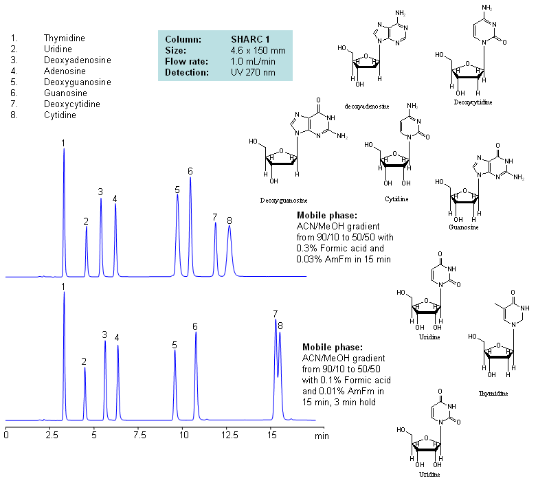
| Column | Sharc 1, 4.6×150 mm, 5 µm, 100A |
| Mobile Phase | MeCN/MeOH |
| Buffer | AmFm, Formic acid |
| Flow Rate | 1.0 ml/min |
| Detection | UV, 270 nm |
| Class of Compounds |
Drug, Acid, Hydrophilic, Ionizable, Vitamin, Supplements |
| Analyzing Compounds | Thymidine, Uridine, Deoxyadenosine, Adenosine, Deoxyguanosine, Guanosine, Deoxycytidine, Cytidine |
Application Column
SHARC 1
The SHARC™ family of innovative columns represents the first commercially available columns primarily utilizing separation based on hydrogen bonding. SHARC stands for Specific Hydrogen-bond Adsorption Resolution Column. Hydrogen bonding involves an interaction or attraction between a bound hydrogen atom and molecules containing electronegative atoms, such as oxygen, nitrogen, and fluorine.
Select optionsCytidine
Deoxyadenosine
Deoxycytidine
Deoxyguanosine
Guanosine
Thymidine
Uridine
HPLC Separation of Cytidine and Cytosine Using the Hydrogen Bonding Method
June 18, 2012
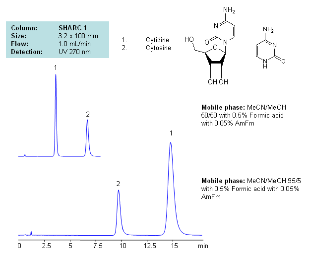
Application Notes: Nucleosides glycosylamines consisting of nucleobase linked to ribose or deoxyribose sugar. Nucleoside are building blocks for DNA and RNA. These compounds are very polar in nature and contain groups available for hydrogen bonding interaction. A method for separation of cytosine and cytidine was developed based on the strong dependence of retention time to the mobile phase composition. The mobile phase consists of acetonitrile and methanol. Order of elution for compounds depends on the amount of acetonitrile and methanol. Our method is compatible with LC/MS and preparative chromatography, and can be used for separation of other nucleobases and nucleotides.
Application Columns: SHARC 1, 3.2×100 mm, 5 um, 100A. To learn more about SHARC 1 columns click here. To order this column click here. To see more chromatographic separations check our web site.
Application Compounds: Cytosine and Cytidine
Detection Technique: UV, LC/MS
| Column | Sharc 1, 3.2×100 mm, 5 µm, 100A |
| Mobile Phase | MeCN/MeOH |
| Buffer | AmFm, Formic acid |
| Flow Rate | 1.0 ml/min |
| Detection | UV, 270 nm |
| Class of Compounds |
Drug, Acid, Hydrophilic, Ionizable, Vitamin, Supplements |
| Analyzing Compounds | Cytidine, Cytosine |
Application Column
SHARC 1
The SHARC™ family of innovative columns represents the first commercially available columns primarily utilizing separation based on hydrogen bonding. SHARC stands for Specific Hydrogen-bond Adsorption Resolution Column. Hydrogen bonding involves an interaction or attraction between a bound hydrogen atom and molecules containing electronegative atoms, such as oxygen, nitrogen, and fluorine.
Select optionsCytosine
HPLC Separation of Thymidine, Uridine, Adenosine, Guanosine, and Cytidine Using the Hydrogen Bonding Method
June 15, 2012
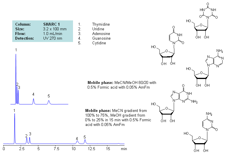
Application Notes: Nucleosides are glycosylamines consisting of nucleobase linked to ribose or deoxyribose sugar and are building blocks for DNA and RNA. These compounds are very polar and contain groups available for hydrogen bonding interaction. Thymidine, uridine, adenosine, guanosine and cytidine were separated using a hydrogen-bonding method. There is a strong correlation between the retention time and mobile phase composition. The strength of hydrogen-bonding interaction increases as the number of hydroxyls in the analytes increase. Additionally the order of elution for compounds depends on the ratio of the mobile phases: acetonitrile and methanol. Our method is compatible with LC/MS and preparative chromatography.
Application Columns: SHARC 1, 3.2×100 mm, 5 um, 100A, To learn more about SHARC 1 columns click here. To order this column click here. To see more chromatographic separations check our web site.
Application Compounds: Thymidine, uridine, adenosine, guanosine and cytidine
| Column | Sharc 1, 3.2×100 mm, 5 µm, 100A |
| Mobile Phase | MeCN/MeOH |
| Buffer | AmFm, Formic acid |
| Flow Rate | 1.0 ml/min |
| Detection | UV, 270 nm |
| Class of Compounds |
Drug, Acid, Hydrophilic, Ionizable, Vitamin, Supplements |
| Analyzing Compounds | Thymidine, Uridine, Adenosine, Guanosine, Cytidine |
Application Column
SHARC 1
The SHARC™ family of innovative columns represents the first commercially available columns primarily utilizing separation based on hydrogen bonding. SHARC stands for Specific Hydrogen-bond Adsorption Resolution Column. Hydrogen bonding involves an interaction or attraction between a bound hydrogen atom and molecules containing electronegative atoms, such as oxygen, nitrogen, and fluorine.
Select optionsCytidine
Guanosine
Thymidine
Uridine
HPLC Separation of Nucleic Bases at pH 4 and 5 on Obelisc N
March 3, 2007
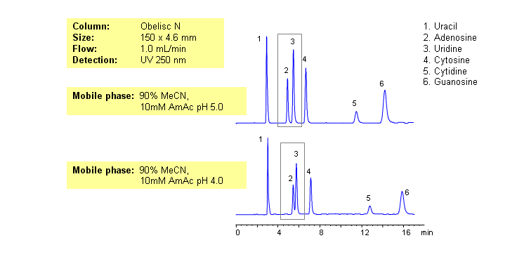
Nucleic bases are biological compounds found in genetic molecules (DNA, RNA). They can be separated on an Obelisc N column, which offers very polar characteristics and can be used with positively or negatively charged groups. Closely-eluted adenosine and uridine can be further separated by simply adjusting the pH of the mobile phase. Mobile phase is water and acetonitrile (MeCN, ACN) with Ammonium Acetate as buffer. UV detection at 250nm.
| Column | Obelisc N, 4.6×150 mm, 5 µm, 100A |
| Mobile Phase | MeCN -90% |
| Buffer | AmAc |
| Flow Rate | 1.0 ml/min |
| Detection | UV, 250 nm |
| Class of Compounds |
Drug, Acid, Hydrophilic, Ionizable, Vitamin, Supplements |
| Analyzing Compounds | Uracil, Uridine, Adenosine, Guanosine, Cytidine, Cytosine |
Application Column
Obelisc N
SIELC has developed the Obelisc™ columns, which are mixed-mode and utilize Liquid Separation Cell technology (LiSC™). These cost-effective columns are the first of their kind to be commercially available and can replace multiple HPLC columns, including reversed-phase (RP), AQ-type reversed-phase, polar-embedded group RP columns, normal-phase, cation-exchange, anion-exchange, ion-exclusion, and HILIC (Hydrophilic Interaction Liquid Chromatography) columns. By controlling just three orthogonal method parameters - buffer concentration, buffer pH, and organic modifier concentration - users can adjust the column properties with pinpoint precision to separate complex mixtures.
Select optionsCytidine
Cytosine
Guanosine
Uracil
Uridine
