HPLC Method for Nicotinamide Adenine Dinucleotide (NAD), Nicotinamide Adenine Dinucleotide (reduced) (NADH) on PEI by SIELC Technologies
High Performance Liquid Chromatography (HPLC) Method for Analysis of SNAD and NADH
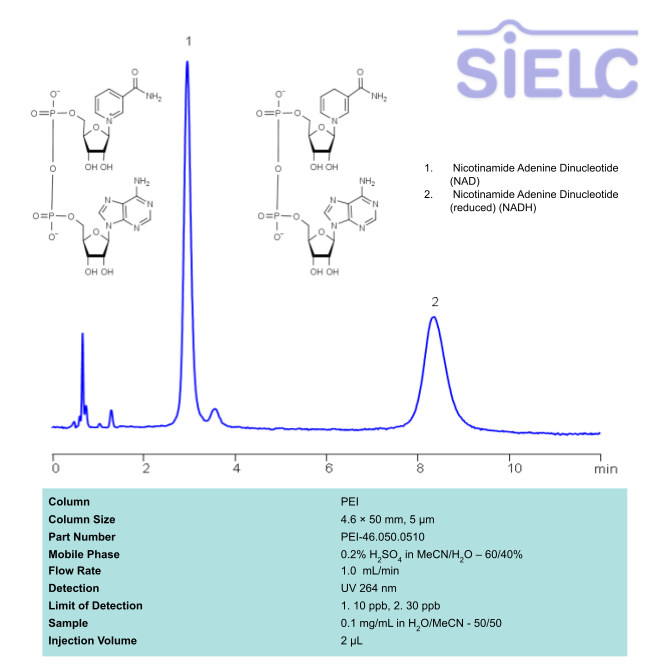
Nicotinamide adenine dinucleotide (NAD), is a coenzyme found in every single living cell. NAD can exist in two forms: NAD+ and NADH. The conversion of NAD from its oxidized form (NAD+) to its reduced form (NADH), and back, provides the cell with a mechanism for accepting and donating electrons.
NAD and NADH can be retained, separated and UV detected at 264 nm using the PEI column with a simple mobile phase of acetonitrile (ACN) and water with sulfuric acid buffer and detected by UV.
| Column | PEI, 4.6 x 50 mm, 5 µm, 100 A, |
| Mobile Phase | MeCN/H2O – 60/40% |
| Buffer | H2SO4 – 0.2% |
| Flow Rate | 1.0 ml/m |
| Detection | UV 264 nm |
| Class of Compounds | Drug |
| Analyzing Compounds | Nicotinamide Adenine Dinucleotide (NAD), Nicotinamide Adenine Dinucleotide (reduced) (NADH) |
Application Column
PEI
Column Diameter: 4.6 mm
Column Length: 50 mm
Particle Size: 5 µm
Pore Size: 100 A
Column options:
Application Analytes:
Nicotinamide Adenine Dinucleotide (NAD)Nicotinamide Adenine Dinucleotide (reduced) (NADH)
Application Detection:
UV Detection
SIELC Technologies usually develops more than one method for each compound. Therefore, this particular method may not be the best available method from our portfolio for your specific application. Before you decide to implement this method in your research, please send us an email to research@sielc.com so we can ensure you get optimal results for your compound/s of interest.