| CAS Number | 635-65-4 |
|---|---|
| Molecular Formula | C33H36N4O6 |
| Molecular Weight | 584.673 |
| InChI Key | BPYKTIZUTYGOLE-UHFFFAOYSA-N |
| LogP | 2.94 |
| Synonyms |
|
Applications:
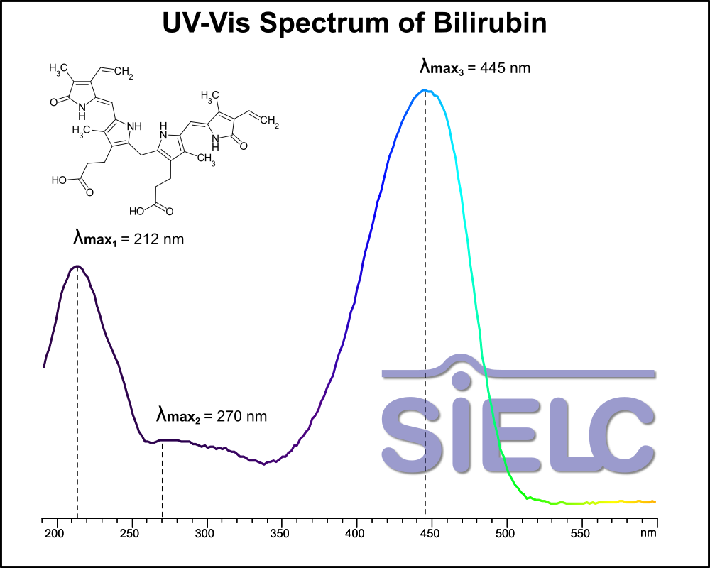
Uv-Vis Spectrum of Bilirubin
February 16, 2026
Access the UV-Vis Spectrum SIELC Library
If you are looking for optimized HPLC method to analyze Bilirubin check our HPLC Applications library
For optimal results in HPLC analysis, it is recommended to measure absorbance at a wavelength that matches the absorption maximum of the compound(s) being analyzed. The UV spectrum shown can assist in selecting an appropriate wavelength for your analysis. Please note that certain mobile phases and buffers may block wavelengths below 230 nm, rendering absorbance measurement at these wavelengths ineffective. If detection below 230 nm is required, it is recommended to use acetonitrile and water as low UV-transparent mobile phases, with phosphoric acid and its salts, sulfuric acid, and TFA as buffers.
For some compounds, the UV-Vis Spectrum is affected by the pH of the mobile phase. The spectra presented here are measured with an acidic mobile phase that has a pH of 3 or lower.

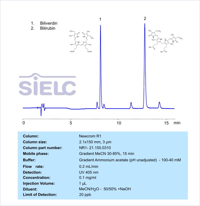
HPLC Method for Separation of Bilirubin and Biliverdin on Newcrom R1 Column
June 23, 2023
HPLC Method for Analysis of Bilirubin, Biliverdin on Newcrom R1 by SIELC Technologies
Separation type: Liquid Chromatography Mixed-mode
Bilirubin and biliverdin are both pigments that are part of the heme degradation pathway in the body. They are structurally related but have different properties and functions.
Bilirubin: This is a yellow compound that is produced by the liver from the breakdown of old red blood cells. It is responsible for the yellow color of bruises and the yellow discoloration seen in conditions such as jaundice and liver disease. Bilirubin is transported in the blood to the liver, where it is conjugated with glucuronic acid and excreted in bile.
Biliverdin: This is a greenish pigment that is an intermediate product in the pathway that produces bilirubin. When red blood cells reach the end of their lifespan (about 120 days), they are broken down in the spleen. The heme group from the hemoglobin in these cells is converted into biliverdin by the enzyme heme oxygenase. This biliverdin is then quickly reduced to bilirubin by biliverdin reductase.
While both these compounds are normal products of metabolism, elevated levels can indicate disease. High levels of bilirubin, for example, can cause jaundice, a condition that results in yellowing of the skin and eyes. Furthermore, since these molecules are breakdown products of heme, they are part of the body’s natural antioxidant defense system. Bilirubin and biliverdin can help protect cells from damage by neutralizing harmful free radicals.
Bilirubin and biliverdin are both part of a class of compounds called tetrapyrroles. More specifically, they belong to a group of tetrapyrroles known as bile pigments.
Tetrapyrroles are a group of organic compounds that contain four pyrrole rings. Heme, found in hemoglobin in red blood cells, is also a type of tetrapyrrole. The breakdown of heme leads to the formation of biliverdin, which is then converted to bilirubin. These bile pigments are then excreted from the body.
Bilirubin and biliverdin can be retained, separate and analyzed on a reverse-phase Newcrom R1, 2.1 x 150 mm, 3 µm, 100 A, dual ended column with a mobile phase consisting of water and Acetonitrile (MeCN) and Ammonium acetate as a buffer. This analysis method can be detected with an Evaporative Light Scattering Detector (ELSD) or any other evaporative detection method (CAD, ESI-MS).
LOD was determined for this combination of instrument, method, and analyte, and it can vary from one laboratory to another even when the same general type of analysis is being performed.
High Performance Liquid Chromatography (HPLC) Method for Analysis of Bilirubin, Biliverdin
Condition
| Column | Newcrom R1, 2.1 x 150 mm, 3 µm, 100 A, dual ended |
| Mobile Phase | Gradient MeCN/H2O -30-85%, 15 min |
| Buffer | Gradient Ammonium acetate (pH unadjusted) – 100-40mM |
| Flow Rate | 0.2 ml/min |
| Detection | UV 405 nm |
| Peak Retention Time | 8.62, 12.8min |
| Sample concentration | 0.1 mg/ml |
| Injection volume | 2 µl |
| Sample diluent | MeCN/H2O -50/50% +NaOH |
| LOD | 20 ppm |
Description
| Class of Compounds | Tetrapyrroles |
| Analyzing Compounds | Bilirubin, Biliverdin |
Application Column
Newcrom R1
Column Diameter: 2.1 mm
Column Length: 150 mm
Particle Size: 3 µm
Pore Size: 100 A
Column options: dual ended
Biliverdin
Separation of Bilirubin on Newcrom R1 HPLC column
May 16, 2018

Bilirubin is a compound that the body produces naturally through the breakdown of red blood cells. It makes urine yellow. Bilirubin can be analyzed by this reverse phase (RP) HPLC method with simple conditions. The mobile phase contains acetonitrile (MeCN), water, and phosphoric acid. For Mass-Spec (MS) compatible applications the phosphoric acid needs to be replaced with formic acid. Smaller 3 µm particle columns are available for fast UPLC applications. This liquid chromatography method is scalable and can be used for isolation of impurities in preparative separation. It also suitable for pharmacokinetics.
.
Application Column
Newcrom R1
The Newcrom columns are a family of reverse-phase-based columns. Newcrom A, AH, B, and BH are all mixed-mode columns with either positive or negative ion-pairing groups attached to either short (25 Å) or long (100 Å) ligand chains. Newcrom R1 is a special reverse-phase column with low silanol activity.
Select options